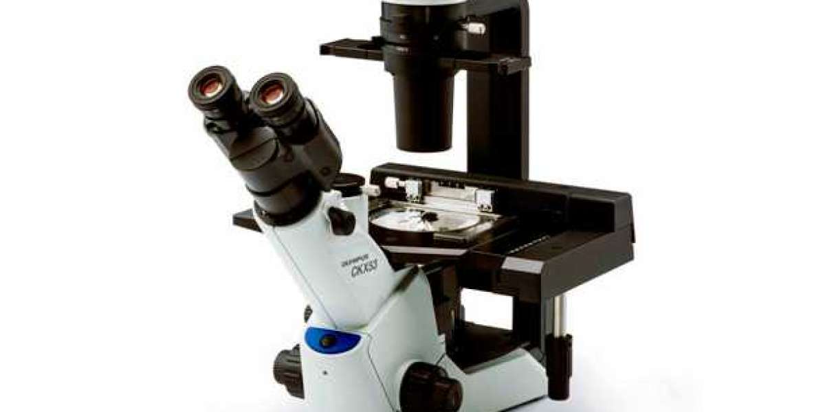
Illumination System in an Inverted Microscope
An inverted microscope is a valuable tool used in various scientific and research fields, particularly in the field of life sciences. One of its critical components is the illumination system, which plays a key role in providing adequate and controlled light to the specimen being observed.

Components of the Illumination System
The illumination system in an inverted microscope consists of several components designed to deliver optimal light to the specimen. These components include:
- Light Source: In most modern inverted microscopes, the light source is typically an LED (Light Emitting Diode) or a halogen lamp. The choice of light source can significantly impact the quality of illumination and the lifespan of the system.
- Condenser: The condenser serves to focus and control the light before it reaches the specimen. It often includes adjustable diaphragms to regulate the angle and size of the light beam, allowing for optimal illumination.
- Filters: Various filters, such as neutral density filters and color filters, may be employed to modify the quality and color of the light reaching the specimen. These filters enable researchers to enhance contrast and reduce glare during observation.
- Beam Splitter: In some advanced inverted microscopes, a beam splitter may be utilized to direct light to different components of the microscope, such as the camera for imaging or additional detectors for specialized techniques.
Role of the Illumination System
The illumination system in an inverted microscope serves several crucial roles in ensuring high-quality imaging and observation:
- Enhancing Contrast: Through the controlled delivery of light, the illumination system enables researchers to enhance contrast and highlight specific features within the specimen, making it easier to visualize and analyze.
- Minimizing Photobleaching: Properly controlled illumination can help reduce the risk of photobleaching, a phenomenon where prolonged or excessive light exposure causes fading or damage to the specimen, particularly in fluorescence microscopy.
Supporting Specialized Techniques: Advanced illumination systems are essential for techniques such as phase contrast, differential interference contrast (DIC), and fluorescence microscopy, where precise control and manipulation of light are critical for successful imaging.
- Illumination System in an Inverted Microscope
Advanced Illumination Techniques
Inverted microscopes often employ advanced illumination techniques to cater to specific research and imaging requirements:
- Fluorescence Illumination: This technique utilizes specific wavelength excitation light to induce fluorescence in labeled specimens. A combination of filters, dichroic mirrors, and a powerful light source is employed to achieve this type of illumination, enabling researchers to visualize cellular structures and molecular interactions.
- Phase Contrast Illumination: Phase contrast microscopy utilizes special optical components, including phase plates and annular diaphragms, to convert phase shifts in light passing through a transparent specimen into intensity variations. This technique provides detailed imaging of transparent, unstained specimens without the need for traditional staining methods.
Darkfield Illumination: Darkfield microscopy utilizes oblique or annular illumination to selectively illuminate the specimen from the side. This method is effective for enhancing contrast and revealing fine details in highly light-scattering specimens, such as live cells, bacteria, or subcellular structures.
The Role of Inverted Microscopes in Various Fields
Inverted microscopes equipped with sophisticated illumination systems are extensively used in diverse scientific and research fields:
- Cell Biology: In cell biology, inverted microscopes facilitate the visualization of living cells, cell cultures, and subcellular structures using advanced techniques such as fluorescence and time-lapse imaging.
- Microbiology: In microbiology, these microscopes are instrumental for observing bacteria, fungi, and other microorganisms. Techniques like darkfield illumination aid in visualizing these specimens with enhanced contrast.
Neuroscience: In neuroscience research, inverted microscopes play a crucial role in imaging neural cultures, neural tissues, and dynamic cellular processes. Fluorescence imaging techniques are particularly valuable in studying neural connectivity and activity.
The illumination system is a fundamental component of an inverted microscope, enabling researchers to achieve high-quality imaging, enhance contrast, and employ advanced techniques for various scientific and research applications. As these microscopes continue to evolve, their illumination systems will play a pivotal role in supporting cutting-edge research and imaging capabilities across the life sciences and material sciences.